MIT researchers mapped activity in individual dendritic spines and somas of visual‑cortex neurons, uncovering five organizing principles – proximity to the soma, micro‑clustering, dendrite type, orientation selectivity and input density – that determine which synapses drive visual responses. The findings provide a quantitative baseline for studying visual disorders and for building more realistic neural network models.
Neurons Follow Simple Spatial Rules to Organize Visual Input in Mouse Cortex
David Orenstein, The Picower Institute for Learning and Memory
Published May 21, 2026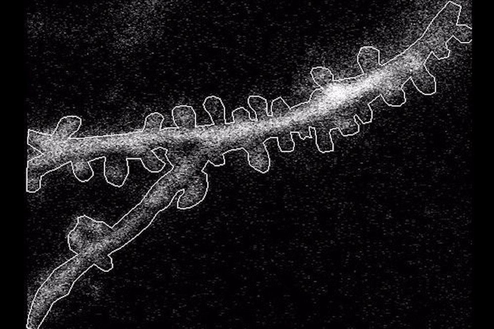
Research breakthrough
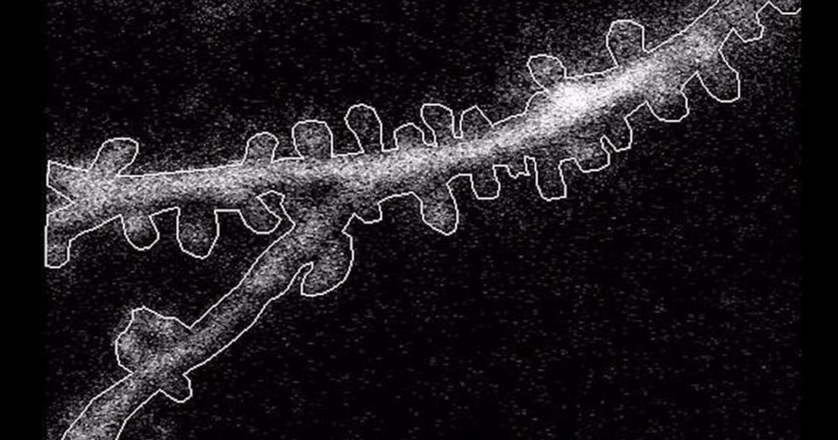
A team led by senior author Mriganka Sur and post‑doc Kyle Jenks published an open‑access paper in iScience that directly visualizes how individual synapses on dendritic spines contribute to the firing of layer 2/3 excitatory neurons in mouse primary visual cortex (V1). By genetically encoding calcium‑sensitive fluorescent reporters in both the soma and each spine, the researchers recorded activity while mice viewed drifting black‑and‑white gratings of different orientations.
Technical approach
- Two‑color calcium imaging – Separate fluorophores labeled the soma and the spine population, allowing simultaneous measurement of somatic action‑potential proxies and local synaptic activation.
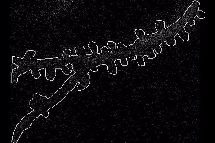
- High‑speed two‑photon microscopy – The setup captured rapid calcium transients across entire dendritic arbors, preserving sub‑micron spatial resolution.
- Quantitative spine‑soma correlation analysis – For each of the 22 recorded neurons (11 visually responsive, 11 unresponsive) the authors computed Pearson correlations between spine activity and somatic calcium traces, then grouped spines by distance, branch type, and orientation selectivity.
- Statistical modeling – A mixed‑effects regression identified which anatomical or functional variables best predicted the strength of spine‑soma coupling.
Five organizing rules uncovered
| Rule | What the data show | Why it matters |
|---|---|---|
| 1. Proximity matters | Spines within ~20 µm of the soma exhibit the strongest correlation with somatic firing; the back‑propagating somatic signal is also detectable only in this inner zone. | Suggests that electrical attenuation limits the influence of distal inputs, shaping how neurons integrate visual cues. |
| 2. Local micro‑clustering | Spines that lie within a 5 µm window tend to fire together, forming “functional enclaves.” Outside this window, co‑activity drops below chance. | Creates discrete processing modules on a single dendrite, potentially sharpening orientation tuning. |
| 3. Apical vs. basal differences | Basal dendrites receive more raw visual drive, but apical dendrites on responsive neurons host a higher proportion of orientation‑selective spines than those on non‑responsive cells. | Highlights that long‑range apical inputs can modulate visual processing when they align with somatic preferences. |
| 4. Orientation selectivity dominates | A spine’s preference for a particular grating orientation explains the largest fraction of variance in its correlation with the soma, far outweighing distance or branch type. | Indicates that functional matching, not just anatomical proximity, is the primary determinant of effective synaptic influence. |
| 5. Global versus local rules | While the above rules operate locally, the overall distribution of responsive spines is non‑random across the whole dendritic tree, reflecting coordinated circuit‑level organization. | Provides a baseline pattern against which disease‑related wiring defects can be compared. |
Real‑world applicability
1. Benchmark for disease models
Genetic mutations linked to autism, schizophrenia, or congenital blindness often disrupt synaptic placement. The quantitative rules from this study give researchers a concrete reference to detect subtle wiring abnormalities in mouse models of these conditions.
2. Improving biologically inspired AI
Current deep‑learning architectures treat each artificial neuron as a point processor. Incorporating dendritic micro‑clusters and distance‑dependent weighting could yield networks that require fewer parameters while preserving orientation selectivity, bringing artificial vision systems closer to cortical efficiency.
3. Guiding neuroprosthetic design
Understanding how somatic signals back‑propagate to shape spine activity informs the placement of stimulation electrodes for visual prostheses. Targeting regions within the 20 µm “effective zone” may maximize the impact of electrical cues on downstream perception.
Limitations and next steps
- Species and layer specificity – The work focuses on mouse V1 layer 2/3; deeper layers or primate cortex may follow different rules.
- Calcium as a proxy – Calcium transients lag behind actual voltage spikes, so the precise timing of synaptic influence remains approximated.
- Static visual stimuli – Only drifting gratings were used; natural scenes with richer statistics could reveal additional organizational principles.
Future experiments plan to (a) extend imaging to awake, behaving mice navigating virtual environments, (b) manipulate specific spine clusters with optogenetics to test causality, and (c) compare the observed rules with large‑scale connectomics data from electron‑microscopy reconstructions.
Broader impact
By quantifying how dendritic architecture and functional selectivity intertwine, this study bridges a gap between microscopic synaptic physiology and macroscopic visual perception. The rules it uncovers will serve as a reference for neuroscientists probing visual disorders, for engineers crafting more efficient neural‑network hardware, and for clinicians developing targeted neurostimulation therapies.
Funding: NIH, Simons Foundation Autism Research Initiative, Freedom Together Foundation. Related resources:
Comments
Please log in or register to join the discussion