MIT researchers have developed a novel nuclear magnetic resonance technique to visualize the disordered outer layer of Tau proteins, a key structure in Alzheimer's disease. This breakthrough reveals how the protein's dynamic 'fuzzy coat' wraps around its rigid core, providing a new roadmap for designing drugs that can penetrate this barrier to stop neurodegenerative tangles.
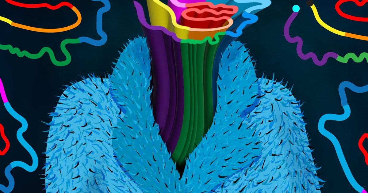
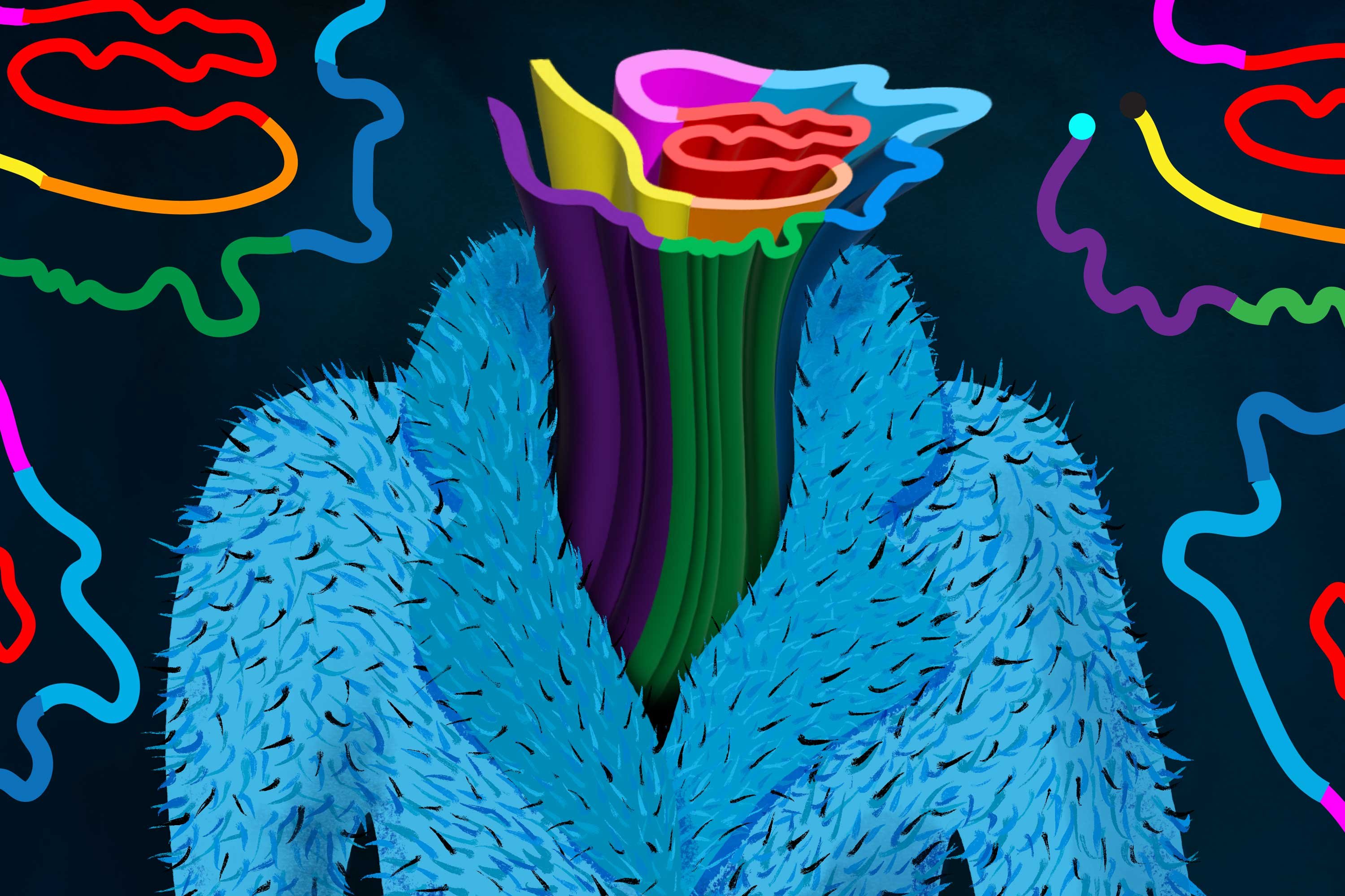
One of the most frustrating challenges in neurodegenerative disease research has been studying the full structure of Tau proteins. While the disease process is driven by the clumping of these proteins into rigid fibrils, about 80 percent of the Tau structure consists of a disordered, constantly shifting outer layer that scientists have been unable to visualize. This 'fuzzy coat' isn't just biological noise—it actively determines how Tau interacts with other molecules and likely dictates how tangles grow in the brain.
MIT chemists have now overcome this barrier by developing a specialized nuclear magnetic resonance (NMR) spectroscopy technique that can capture both the rigid core and the dynamic outer segments of full-length Tau fibrils. Their findings, published in the Journal of the American Chemical Society, provide the first complete structural model of the Tau protein in its pathological state.
The Visualization Problem
In its healthy state, Tau proteins serve a crucial structural role, stabilizing microtubules that act as cellular highways. However, when Tau misfolds, it forms insoluble fibrils characterized by a rigid inner core of beta-sheet strands surrounded by disordered segments. This structural transformation is central to Alzheimer's disease, frontotemporal dementia, and other neurodegenerative conditions.
The problem for researchers has been technical: standard structure determination methods like cryoelectron microscopy and X-ray crystallography excel at capturing static, ordered structures but fail when proteins are highly mobile. The fuzzy coat's constant motion renders these techniques nearly useless for visualizing it.
"If you want to disaggregate these Tau fibrils with small-molecule drugs, then these drugs have to penetrate this fuzzy coat," explains Mei Hong, MIT professor of chemistry and the study's senior author. "Understanding this barrier is essential for developing effective therapeutics."
A New NMR Approach
Hong's team, led by graduate student Jia Yi Zhang, developed an NMR methodology that tracks magnetization transfer between rigid and mobile regions of the protein. Their approach works by first magnetizing protons within the most rigid amino acids, then measuring how long it takes for this magnetization to spread to the mobile segments.
This technique essentially maps the spatial relationships between different parts of the protein by observing how energy moves between them. When magnetization transfers quickly between two regions, it indicates they are physically close. When transfer is slow or absent, they are distant or isolated.
The researchers complemented this with measurements of amino acid mobility, creating a comprehensive picture of both structure and dynamics. "We have now developed an NMR-based technology to examine the fuzzy coat of a full-length Tau fibril, allowing us to capture both the dynamic regions and the rigid core," Hong says.

The Burrito Model
The complete structural model reveals that the Tau protein resembles a burrito, with the rigid core wrapped in multiple layers of the fuzzy coat. The researchers identified three distinct categories of protein segments based on their mobility:
- The rigid core: Beta-sheet strands that form the structural backbone of the fibril
- Intermediate mobility segments: Regions directly surrounding the core that show restricted but measurable movement
- Highly dynamic outer layers: The most mobile segments, particularly rich in the amino acid proline
One surprising finding involved the proline-rich regions. Scientists had previously assumed these positively charged segments would be partially immobilized by their proximity to the rigid core's positive charges. Instead, the NMR data showed they are highly mobile, suggesting electrostatic repulsion keeps them in constant motion.
This dynamic structure has important implications for how Tau fibrils grow. The burrito-like wrapping indicates that normal Tau proteins are more likely to add onto the ends of existing filaments rather than piling onto the sides. This growth mechanism differs from other protein aggregation models and could influence how therapeutics interrupt the process.
Practical Implications for Drug Development
The ability to visualize the fuzzy coat directly addresses a major pharmaceutical challenge. Most drug candidates designed to block Tau aggregation have failed because they cannot effectively penetrate the disordered outer layer to reach their target sites on the rigid core.
With this new structural map, researchers can:
- Design molecules that specifically target the dynamic regions to destabilize the fibril structure
- Identify binding sites on the fuzzy coat that could serve as therapeutic targets
- Understand how existing drug candidates interact with both the core and outer layers
- Model how mutations that alter the fuzzy coat affect aggregation propensity
The research team plans to use their technique to study how misfolded Tau proteins from Alzheimer's patients can serve as templates to convert normal Tau into the pathological form. This could reveal critical vulnerabilities in the aggregation cascade.
The work was funded by the National Institutes of Health and represents a significant methodological advance that could accelerate drug discovery for Alzheimer's disease and related neurodegenerative conditions.
Paper: "Heterogeneous Dynamics of the Fuzzy Coat of Full-Length Phospho-Mimetic Tau Fibrils" - Journal of the American Chemical Society
Related Research: Hong Lab at MIT Department of Chemistry
Comments
Please log in or register to join the discussion