MIT researchers discovered a shared brain wave pattern in humans and mice with fragile X syndrome, providing a crucial biomarker for evaluating neurological treatments.
Researchers at MIT's Picower Institute for Learning and Memory have identified a consistent brain wave pattern in fragile X syndrome that bridges human patients and mouse models, potentially revolutionizing treatment evaluation for neurological disorders. This discovery addresses a persistent challenge in translational neuroscience: therapies showing promise in animal models often fail in human trials due to the lack of objective, comparable physiological markers.
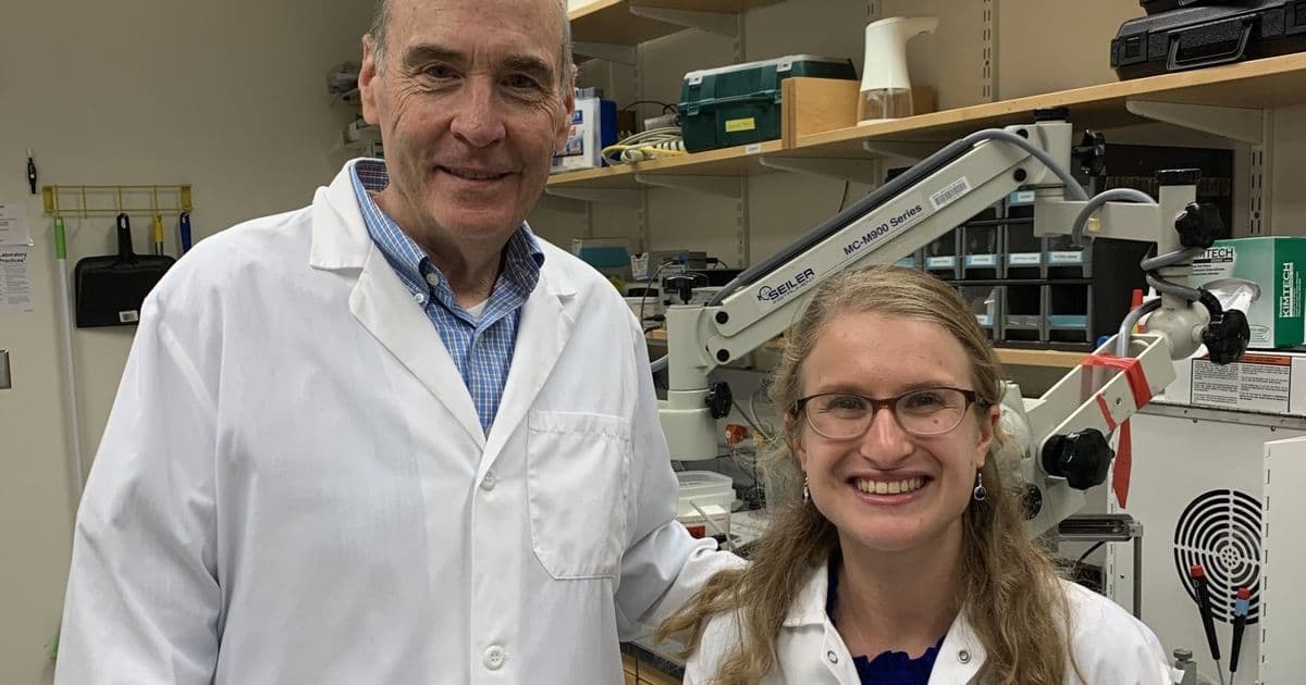
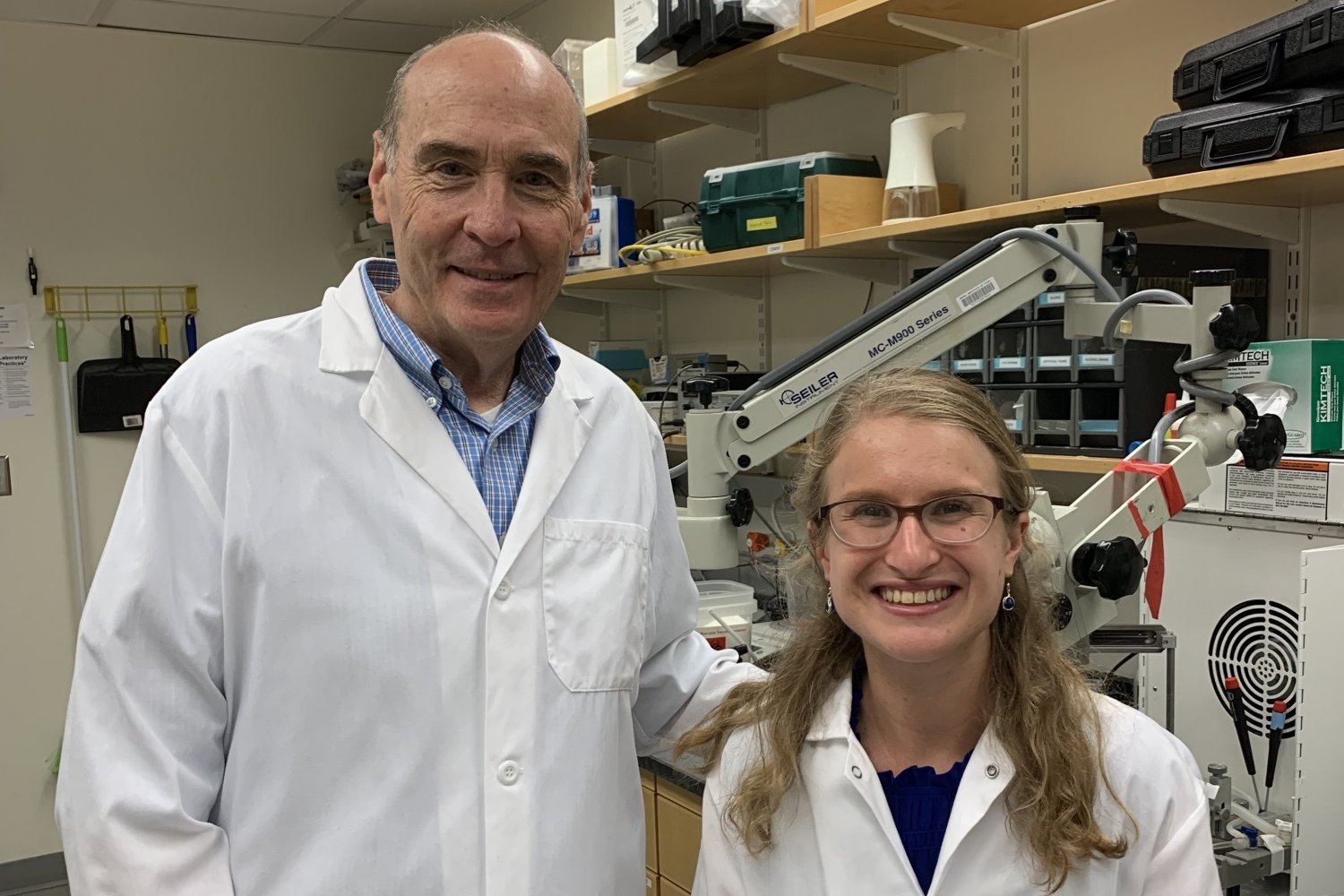
Led by postdoctoral researcher Sara Kornfeld-Sylla and Picower Professor Mark Bear, the team analyzed electroencephalogram (EEG) recordings from boys and men with fragile X syndrome alongside age-matched neurotypical individuals. Simultaneously, they measured brain activity in mice genetically modified to model the disorder. Using a novel analytical approach that isolates periodic fluctuations in brain wave power across the frequency spectrum, the researchers uncovered species-spanning patterns.
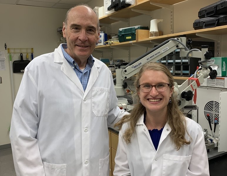
Mark Bear (left) and Sara Kornfeld-Sylla led the study identifying a fragile X biomarker shared between species. "This research weaves together different datasets and finds the connection between brain wave activity in fragile X humans and mouse models," Kornfeld-Sylla noted.
The key biomarker manifests in low-frequency brain waves (theta band in mice, alpha band in humans) and exhibits two distinct signatures:
- Adults: A significant shift in peak power frequency toward slower waves
- Juveniles: Marked reduction in power at the characteristic peak frequency
Further analysis revealed these patterns originate from two subpeaks within the low-frequency band, with the lower subpeak specifically altered in fragile X cases. Through neural circuit experiments in mice, the team traced this biomarker to impaired function of somatostatin-expressing interneurons—inhibitory neurons that shape brain wave patterns via the neurotransmitter GABA.
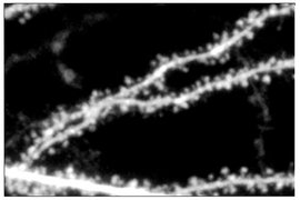
Micrograph showing neural dendrites, where impaired GABA signaling contributes to the observed brain wave alterations in fragile X syndrome.
The biomarker's clinical relevance was validated through pharmacological testing with arbaclofen—a candidate therapy that enhances GABA signaling. Single doses corrected the biomarker in juvenile fragile X mice, with effects scaling with dosage. Notably, neurotypical mice responded to lower doses than fragile X models, reflecting the latter's reduced GABA receptivity.
"Identifying this electrophysiological signature allows direct cross-species comparison of treatment effects," explained Bear. "We can now ask: At what dose does a drug alter this signature similarly in mice and humans? This creates a translational bridge between physiological measures and behavioral outcomes."
The biomarker's discovery emerged from collaborative data sharing across seven institutions, including Boston Children's Hospital and King's College London. Its significance extends beyond fragile X syndrome, as similar low-frequency brain wave disruptions appear in multiple neurological conditions. This approach could accelerate treatment development for autism spectrum disorders by providing an objective, non-invasive measure of therapeutic impact.
Funding was provided by the National Institutes of Health, National Science Foundation, FRAXA Research Foundation, and several private foundations supporting autism research.
Comments
Please log in or register to join the discussion