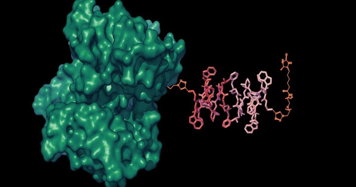
A new class of liposomal nanoparticle reporters (LisNRs) developed at MIT can brighten or dim MRI contrast in response to specific molecules, offering up to ten‑fold sensitivity gains over traditional agents and opening a path toward brain‑wide imaging of neurotransmitters.
MIT engineers unveil liposomal nanoparticle reporters that amplify MRI signals for molecular imaging
Magnetic resonance imaging (MRI) has long been prized for its ability to render high‑resolution maps of anatomy and blood flow without ionising radiation. Yet the technique has struggled to detect low‑abundance neurochemicals because conventional contrast agents require a one‑to‑one interaction with each target molecule, limiting the observable signal. In a paper published in Nature Biomedical Engineering (May 13, 2026), Alan Jasanoff’s group at MIT reports a fundamentally different approach: liposomal nanoparticle reporters (LisNRs) that let a single molecular event modulate many gadolinium‑based contrast agents, dramatically amplifying the MRI readout.
Technical approach: water‑gated liposomal contrast
LisNRs are built from three core components:
- A liposomal shell – a nanoscale vesicle (~100 nm) that encloses thousands of gadolinium chelates. Inside the vesicle the gadolinium is magnetically isolated from bulk water, so it does not affect the MRI signal.
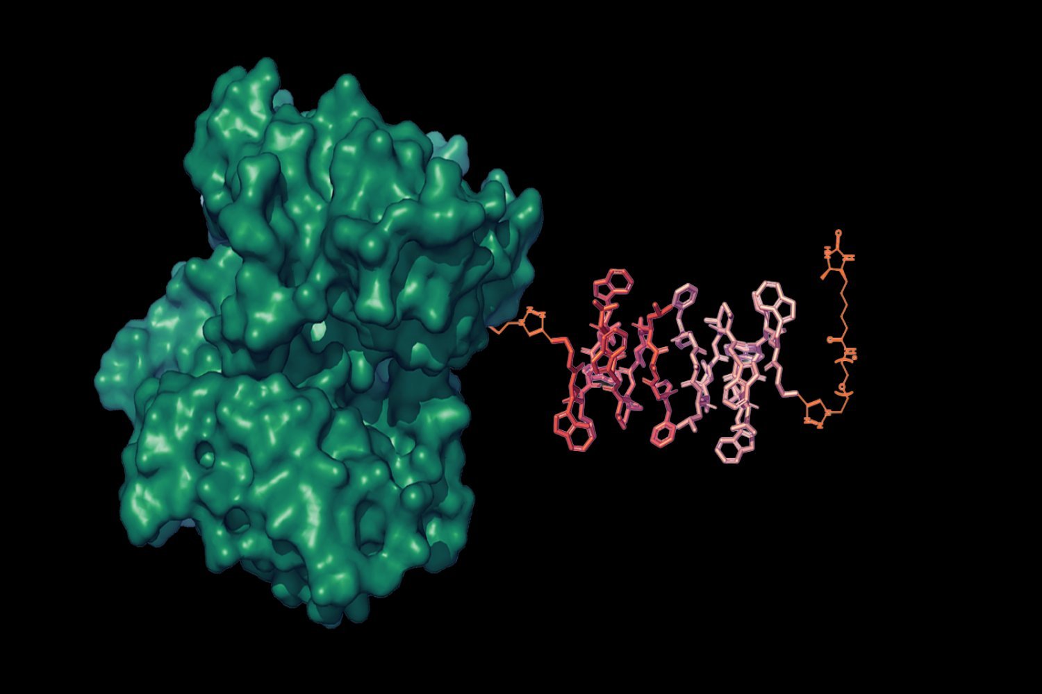
- Engineered water channels – protein pores (derived from aquaporin or synthetic designs) inserted into the liposome membrane. Their opening is controlled by a molecular “lock” that responds to a specific target.
- A blocking protein or peptide – a small binder that occupies the channel pore until the target molecule displaces it (or, for the inverse design, the target itself blocks an otherwise open channel).
When the target molecule is present, the lock is removed and water floods the interior of the liposome. The high concentration of gadolinium now interacts with the surrounding water protons, increasing the longitudinal relaxation rate (T1) and producing a brighter voxel in the MR image. The reverse design produces a dimming effect by sealing the channel.
Because each liposome carries on the order of 10⁴–10⁵ gadolinium ions, a single target‑binding event can toggle the contrast of many agents simultaneously. This “amplification” translates into an order‑of‑magnitude improvement in detection limit compared with traditional small‑molecule probes that require a stoichiometric ratio of contrast agent to target.
Design workflow
- Target selection – The team first identifies a ligand that can be recognised by a high‑affinity protein or aptamer.
- Channel engineering – Using computational protein design, they graft a binding pocket onto an aquaporin scaffold so that ligand binding induces a conformational change that opens the pore.
- Liposomal formulation – Standard thin‑film hydration and extrusion methods generate uniform vesicles; gadolinium‑DTPA is encapsulated at high loading, and the engineered channels are reconstituted into the membrane.
- In‑vitro validation – Fluorescence‑based water‑flux assays confirm that channels open only in the presence of the target.
- In‑vivo testing – Systemic injection into rats followed by T1‑weighted MRI demonstrates brightening in regions where the target (e.g., biotin) accumulates.
Real‑world applicability and early results
In the first demonstration, the researchers packaged a biotin‑responsive LisNR and injected it into live rats. Biotin, a small vitamin that can be delivered systemically, served as a proof‑of‑concept target. MRI scans showed a clear, localized increase in signal intensity at biotin‑rich sites, with a reported ten‑fold sensitivity improvement over a conventional gadolinium chelate that relies on direct binding.
Key practical advantages observed in the study:
- Systemic delivery – The liposomes crossed the blood‑brain barrier in sufficient quantities to generate detectable contrast throughout the brain, suggesting that peripheral administration could be viable for many neurochemical studies.
- Reversibility – By swapping the blocking protein for a target‑dependent blocker, the same platform can be tuned to either brighten or dim signals, giving experimenters flexibility in experimental design.
- Scalability – The liposomal formulation uses well‑established pharmaceutical manufacturing steps (extrusion, sterilisation), easing the path toward pre‑clinical translation.
Limitations to address
While the results are promising, several challenges remain before LisNRs can be used to monitor endogenous neurotransmitters such as dopamine or glutamate:
- Target affinity – Many neurochemicals are present at sub‑micromolar concentrations. The binding domain must achieve nanomolar affinity while still allowing rapid on/off kinetics.
- Temporal resolution – Water exchange through the channel is fast, but the overall MRI acquisition time limits detection of rapid neurotransmitter spikes. Combining LisNRs with ultrafast imaging sequences (e.g., magnetic resonance fingerprinting) could mitigate this.
- Safety and clearance – Gadolinium‑based agents have raised concerns about deposition in the brain. Encapsulation inside liposomes reduces free gadolinium exposure, but long‑term biodistribution studies are needed.
- Immunogenicity – The engineered protein channels and blocking peptides could provoke immune responses. Humanising the protein scaffolds and employing stealth coatings (PEGylation) are standard strategies to minimise this risk.
Outlook: from molecular reporters to brain‑wide chemical mapping
The LisNR platform establishes a modular toolbox: swapping the ligand‑binding domain changes the target, while the same liposomal chassis and water‑gate architecture remain constant. The Jasanoff lab plans to target dopamine and glutamate next, leveraging the high abundance of these transmitters to validate the approach under physiologically relevant conditions.
If successful, LisNRs could complement existing functional MRI (fMRI) methods that rely on hemodynamic changes, providing a direct readout of chemical signaling rather than an indirect vascular proxy. Such capability would enable researchers to map the spatiotemporal dynamics of neuromodulators across the whole brain, a long‑standing goal in systems neuroscience.
Funding and collaborations – The work was supported by the Lore Harp McGovern Fund, the Yang Tan Collective, and several MIT‑based centers focused on brain‑body research. Collaboration with Masayuki Inoue’s group at the University of Tokyo supplied high‑potency engineered channels, illustrating the interdisciplinary nature of the project.
For a deeper dive into the methodology, the open‑access version of the paper can be found on the journal’s website.
Comments
Please log in or register to join the discussion