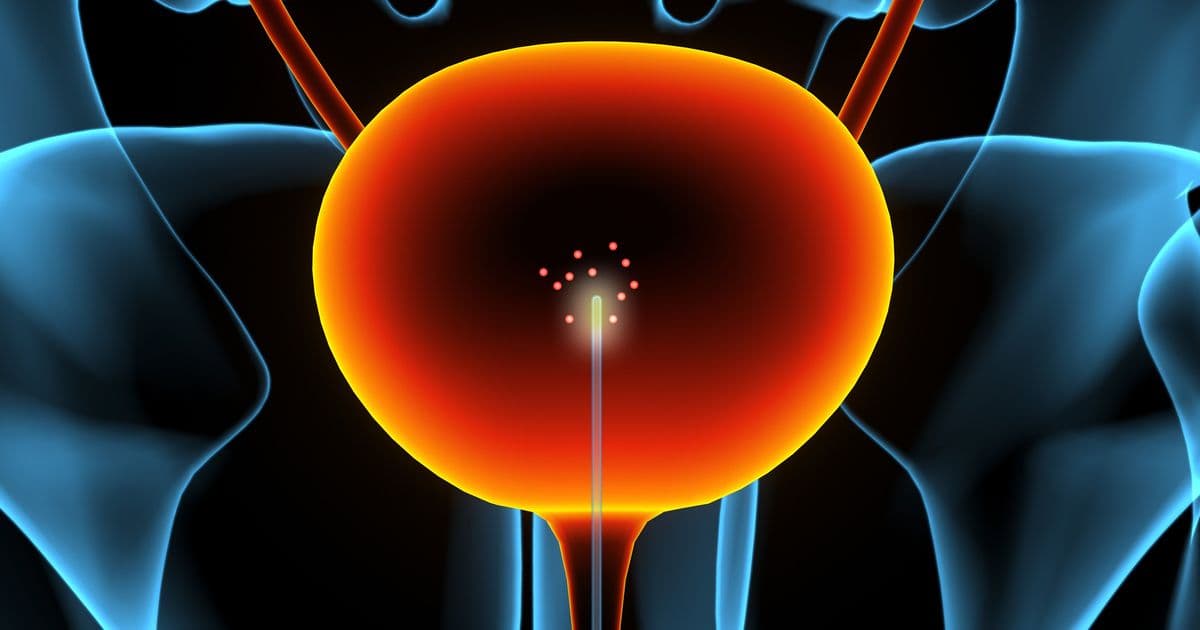
MIT researchers have built a catheter coated with carbon‑nanotube nanosensors that can locate the bladder‑cancer biomarker NMP‑22 in situ, achieving sensitivity orders of magnitude beyond standard urinalysis and enabling chemical imaging of tumors as small as 16 mm².
Carbon‑nanotube catheter offers molecular imaging for earlier bladder‑cancer detection
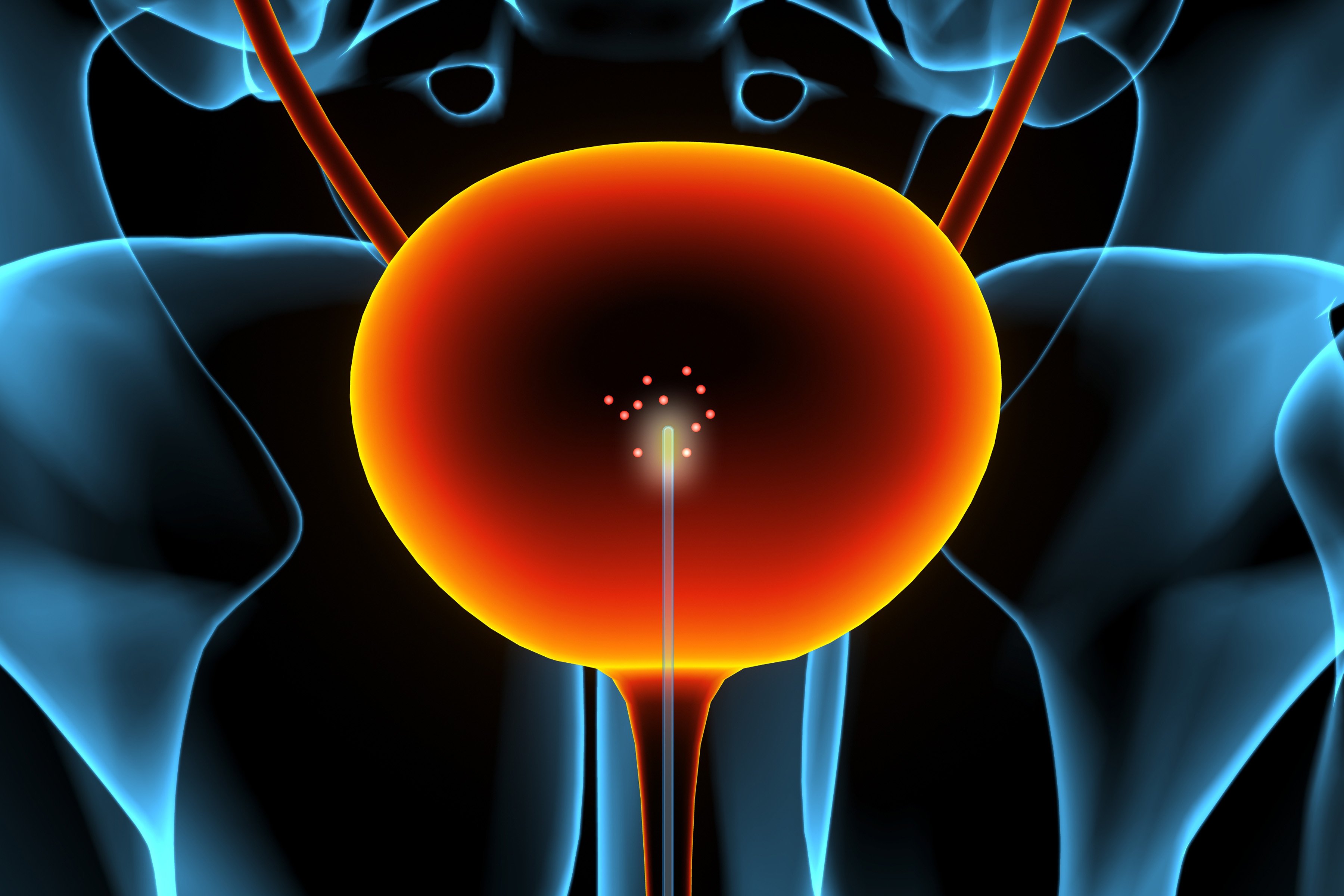
Bladder cancer recurs in roughly half of patients within five years of treatment, making timely monitoring a costly clinical challenge. A team led by MIT’s Michael Strano has demonstrated a new approach that brings the sensor to the disease site, using a catheter whose inner surface is coated with carbon‑nanotube arrays that fluoresce in response to a cancer‑associated protein.
Technical approach
The device relies on single‑walled carbon nanotubes (SWCNTs) that emit near‑infrared light when excited by a laser. By wrapping each nanotube with a synthetic polymer that binds specifically to nuclear matrix protein 22 (NMP‑22), the researchers created a sensor whose fluorescence wavelength or intensity shifts when the target protein is present. The catheter tip incorporates a tiny rotating ball lens that both delivers the excitation laser and collects the emitted fluorescence. As the lens sweeps 360°, the system records the color and intensity of each signal, allowing it to reconstruct a spatial map of NMP‑22 concentration along the bladder wall.
Key engineering details:
- Nanotube functionalization – Polymers are tuned to present binding sites that recognize NMP‑22 with high affinity, ensuring that even picomolar concentrations trigger a measurable optical change.
- Optical readout – Near‑infrared fluorescence penetrates tissue better than visible light, reducing background scattering and enabling detection through the thin bladder lining.
- Signal processing – Custom algorithms convert wavelength shifts into concentration estimates and assign each measurement to a specific angular position of the lens, producing a “chemical image” of the bladder interior.
In a rodent model, the system detected NMP‑22 at levels roughly 50,000 times lower than conventional urine tests and pinpointed tumor locations as small as 16 mm². The sensitivity gain stems from measuring the biomarker directly at its source, before dilution and enzymatic degradation in the urine.
Real‑world applicability
Current surveillance relies on periodic cystoscopy and urine‑based assays. Cystoscopy provides visual confirmation but is invasive and may miss sub‑urothelial lesions; urine tests are cheap but suffer from low specificity and delayed detection. The nanotube‑sensor catheter bridges this gap by offering:
- Earlier detection – Molecular signals appear before a tumor breaches the urothelium, potentially allowing clinicians to intervene when lesions are still microscopic.
- Targeted biopsies – The chemical image can guide a physician to the exact region of elevated biomarker, reducing the number of blind biopsies.
- Office‑based workflow – The team is miniaturizing the optics and electronics so the procedure could be performed during a routine office visit, without the need for a full endoscopic suite.
Beyond bladder cancer, the platform is adaptable. By swapping the polymer coating, the same catheter could sense biomarkers for gastrointestinal, cardiovascular, or infectious diseases. Strano’s group has already demonstrated nanotube sensors for hydrogen peroxide, riboflavin, and viral proteins, suggesting a modular toolbox for endoscopic diagnostics.
Limitations and next steps
While the proof‑of‑concept results are compelling, several practical hurdles remain:
- Regulatory pathway – The catheter combines a medical device with a novel nanomaterial; safety testing will need to address long‑term biocompatibility of carbon nanotubes in the urinary tract.
- Manufacturing scale – Uniform coating of nanotube arrays on flexible catheter surfaces must be reproducible at commercial volumes.
- Signal robustness – Variations in bladder geometry, urine composition, and patient movement could affect optical readout; robust calibration routines will be essential.
The researchers are now engineering a compact imaging module that can be detached from the catheter and sterilized separately, and they are testing the system in larger animal models that more closely mimic human bladder anatomy.
If these challenges are met, the technology could shift bladder‑cancer surveillance from a periodic, invasive visual exam to a rapid, molecular‑level screening that catches recurrence at its earliest stage, reducing treatment costs and improving patient outcomes.
For more details, see the paper “Chemical efflux imaging using an annular nanosensor array for in situ bladder cancer detection” in Nature Nanotechnology and the MIT Strano Group page.
Comments
Please log in or register to join the discussion