MIT researchers have created the first "living" implant that rewires sensory nerves to paralyzed organs, using the body's own tissue as hardware to restore movement and sensation.
What if a paralyzed bladder could empty itself again, or a damaged intestine could push food forward without external assistance? A breakthrough from MIT researchers offers a glimpse into this future with a technology that transforms living muscle into computer-controlled motors capable of reviving organs that have lost their connection to the brain.
In a study published today in Nature Communications, the team introduces the myoneural actuator (MNA) - a biohybrid system that reprograms existing muscles to become fatigue-resistant, implantable motors. The technology represents the first living implant that uses rewired sensory nerves to restore movement in paralyzed organs while simultaneously transmitting sensations back to the brain.
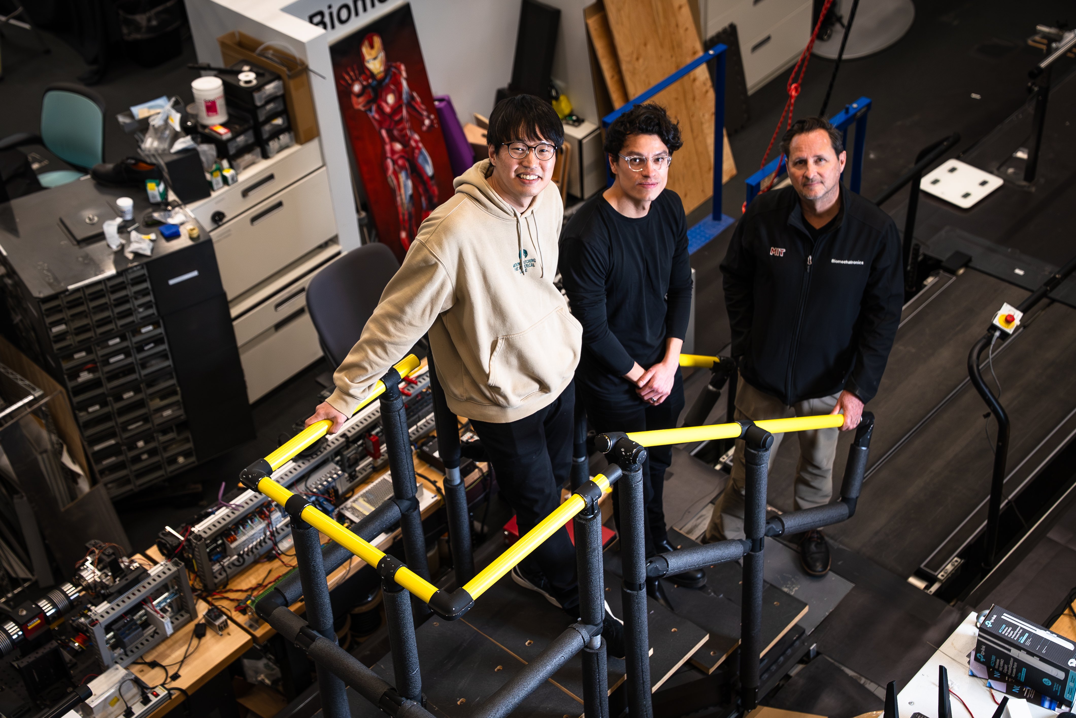
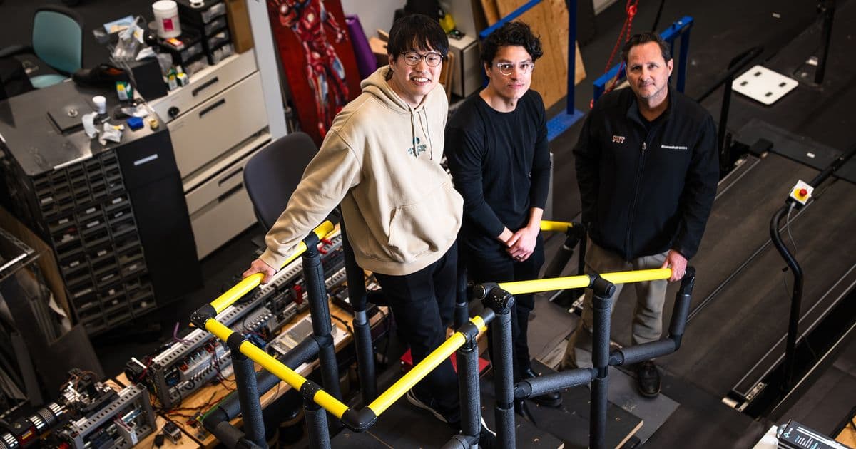
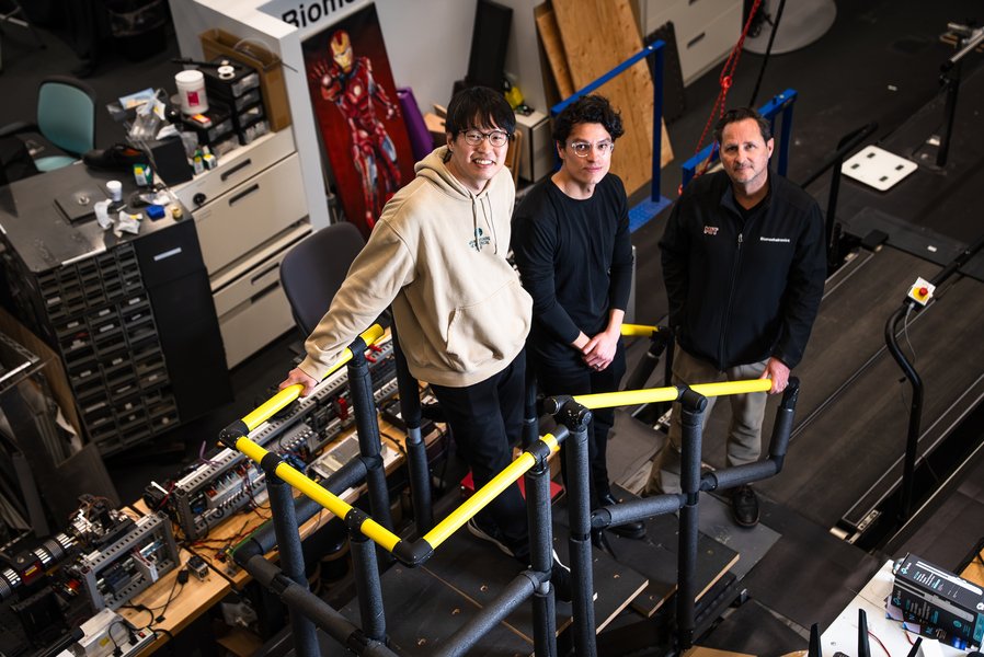
"We've built an interface that leverages natural pathways used by the nervous system so that we can seamlessly control organs in the body, while also enabling the transmission of sensory feedback to the brain," explains Hugh Herr, senior author of the study and professor at the MIT Media Lab. The research was co-led by Herr's postdoc Guillermo Herrera-Arcos and former postdoc Hyungeun Song.
The challenge of restoring organ function has long stymied researchers. Traditional approaches have included miniaturized mechanical actuators or lab-grown muscle tissue, but both approaches face significant limitations. Mechanical actuators struggle to operate efficiently at the centimeter scale required for organ-level control, while tissue engineering remains time-intensive and far from clinical readiness.
Herr's team took a fundamentally different approach: engineering existing muscles to become actuators that can reinstate motion in organs. The key insight was recognizing that sensory neurons, unlike motor neurons, are wired to receive rather than command signals. By rerouting motor signals through sensory fibers, the researchers created a system where a computer - not the brain - becomes the muscle's command center.
"You don't want the brain to consciously control the muscle actuator because you want the actuator to automatically control an organ, like the heart," explains Herrera-Arcos. This approach elegantly bypasses damaged brain pathways while maintaining autonomous organ function.
The discovery that sensory nerves could successfully re-innervate muscle and form functional synapses was particularly remarkable. When the team replaced motor nerves in rodent muscle with sensory ones, the sensory nerves not only integrated but also dramatically improved muscle performance. The sensory-nerve-controlled muscles showed 260 percent greater fatigue resistance compared to native muscles.
This fatigue resistance stems from the fundamental differences between motor and sensory neuron axons. Motor neuron axons vary greatly in size, with larger axons firing first and exhausting the muscle quickly. Sensory axons, however, are nearly uniform in size, allowing signals to broadcast more evenly across muscle fibers and preventing rapid fatigue.
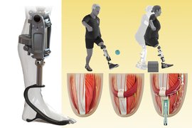
The researchers combined these elements into the myoneural actuator system. In experiments with paralyzed rodent intestines, the MNA successfully reinstated the organ's squeezing motion. The system also controlled rodent calf muscles in experiments designed to mimic residual muscle in human lower-limb amputations.
Critically, the MNA system transmitted sensory signals to the brain, suggesting potential for seamless organ-brain integration. "This suggests that our technology could seamlessly link organs to the brain. For example, we might be able to make a paralyzed stomach relay hunger," explains Song.
The clinical implications are profound. Unlike mechanical devices or organ transplants that introduce foreign materials into the body, MNAs would use the patient's own tissue as hardware. The implantation procedure would leverage existing surgical techniques already commonplace in clinical practice, potentially offering a simpler and safer path to restoring organ function.
Beyond medical applications, the technology opens possibilities for enhancing human-machine interfaces. The researchers envision applications ranging from restoring tactile feedback in prosthetic users to augmenting virtual reality experiences. "The idea is that, if we couple the MNA system to skin and muscles, a person could feel what their virtual avatar is touching even though their real body isn't moving," says Song.
For the millions living with organ dysfunctions, this technology could represent a paradigm shift. "Today's solutions are mostly synthetic: pacemakers and other mechanical assist devices. A living muscle actuator implanted alongside a weakened organ would be part of the body itself. That is a category of medicine different from anything seen in clinic," explains Herrera-Arcos.
Skin applications are particularly intriguing. "Hypothetically, we could wrap MNAs around skin grafts to relay tactile feedback, such as strain or tension, which is currently missing for users of prostheses," Song notes.
While bringing the MNA to clinical use will require extensive testing in larger animal models and eventually human trials, the technology represents a fundamental shift in how we approach organ dysfunction. By turning the body's own tissue into programmable hardware, MIT's researchers have opened a new frontier in biohybrid medicine - one where science fiction becomes medical reality.
The research was funded by the Yang Tan Collective at MIT, K. Lisa Yang Center for Bionics at MIT, Nakos Family Bionics Research Fund at MIT, and the Carl and Ruth Shapiro Foundation.
Comments
Please log in or register to join the discussion