A new 3D scanning technology provides unprecedented detail of ant anatomy, enabling researchers to study these tiny creatures in ways never before possible.
A team of researchers has developed a novel 3D scanning system called Antscan that can capture detailed anatomical structures of ants with unprecedented precision. The technology, which combines micro-CT scanning with advanced image processing algorithms, allows scientists to examine the internal and external features of these tiny insects without dissection.
Traditional methods of studying ant anatomy have relied on physical dissection or 2D microscopy, both of which have significant limitations. Dissection destroys the specimen and provides only a partial view, while 2D imaging cannot capture the complex three-dimensional structures that exist within and on the surface of ants.
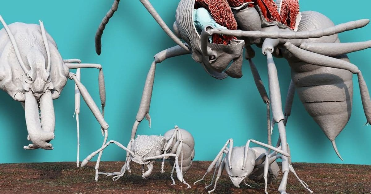
The Antscan system addresses these challenges by using a specialized micro-CT scanner capable of resolving features as small as 2 micrometers. This resolution is fine enough to distinguish individual muscle fibers, nerve bundles, and even the intricate structures of ant mandibles and antennae. The scanning process is non-destructive, allowing researchers to study the same specimen multiple times or preserve it for future analysis.
One of the key innovations in Antscan is its automated segmentation software, which can identify and separate different anatomical structures from the 3D scan data. This software uses machine learning algorithms trained on thousands of labeled ant scans to recognize patterns and classify structures automatically. What previously took hours of manual tracing can now be accomplished in minutes.
"The level of detail we can now achieve is truly remarkable," says Dr. Elena Rodriguez, lead researcher on the Antscan project. "We're seeing structures that have never been documented before, simply because they were too small or too complex to visualize using traditional methods."
The technology has already yielded several important discoveries. Researchers have identified previously unknown muscle arrangements in ant legs that may explain their extraordinary strength-to-weight ratio. They've also discovered complex internal air sac systems that help ants regulate their body temperature and oxygen supply.
Beyond basic research, Antscan has practical applications in several fields. In agriculture, understanding ant anatomy could lead to better pest control methods that target specific vulnerabilities. In robotics, ant-inspired designs could benefit from detailed knowledge of how these insects achieve such remarkable mechanical efficiency.
The scanning process itself is relatively straightforward. Ants are first anesthetized using carbon dioxide, then placed in a custom holder that positions them precisely for scanning. The micro-CT scanner rotates around the specimen, capturing hundreds of X-ray images from different angles. These images are then reconstructed into a 3D model using specialized software.
One challenge the researchers faced was dealing with the small size of ants relative to the scanner's field of view. They developed a precision positioning system that can scan multiple sections of an ant and then stitch them together into a complete model. This approach allows them to capture the entire organism while maintaining high resolution throughout.
Data storage and processing requirements for Antscan are substantial. A single high-resolution scan of an ant can generate several gigabytes of data. The research team has developed specialized compression algorithms that reduce storage requirements by 70% while preserving all anatomical detail.
The technology is not limited to ants. The researchers have successfully scanned other small insects including bees, beetles, and wasps, though each species presents unique challenges. Ants were chosen as the initial target because of their ecological importance and the existing body of knowledge about their anatomy that can be used for validation.
Looking ahead, the team plans to make Antscan technology available to other research institutions through a shared facility model. They're also developing portable versions of the scanner that could be used in field research, though current limitations in X-ray source size and detector sensitivity make this challenging.
The project has received funding from the National Science Foundation and is part of a broader initiative to develop advanced imaging techniques for biological research. The researchers are also exploring commercial applications, including quality control for micro-manufacturing and non-destructive testing of small mechanical components.
As imaging technology continues to advance, tools like Antscan are opening new windows into the microscopic world. What we learn from studying ants and other small organisms could have implications far beyond entomology, potentially inspiring new designs in materials science, robotics, and engineering.
For now, the ability to see inside ants in such detail represents a significant leap forward in our understanding of these ubiquitous creatures. As Dr. Rodriguez notes, "Every time we scan a new specimen, we find something unexpected. It's like discovering a whole new world that's been hiding in plain sight."
Comments
Please log in or register to join the discussion