MIT researchers built an augmented-reality ultrasound system that renders a live 3D model of scanned tissue inside a VR headset, letting novices match expert performance on identification and needle-placement tasks. The work targets one of ultrasound's hardest bottlenecks: the mental gymnastics of reconstructing 3D anatomy from flat 2D slices.
Ultrasound is one of the most accessible imaging tools in medicine, but it carries a cognitive tax that rarely shows up in product specs. A technician sweeps a probe across tissue, watches a stream of 2D cross-sections on a monitor, and has to assemble those slices into a coherent 3D picture inside their own head. MIT researchers have built a system that offloads that reconstruction work to a computer and a headset, and their results suggest the change matters most for the people who have the least training.
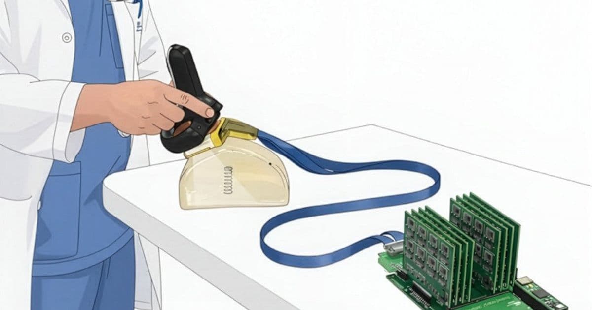
The team, led by associate professor Canan Dagdeviren in MIT's Media Lab, calls the system AR-VIU, for augmented real-time volumetric imaging in ultrasound. Instead of presenting flat images, it renders a live 3D representation of the scanned object and superimposes it over the object's real-world location through an AR/VR headset. The work appears in Nature Communications Engineering, with graduate students Jason Hou and Shrihari Viswanath as lead authors.

The mental tomography bottleneck
To understand why this is useful, it helps to understand what ultrasound actually produces. The probe emits high-frequency sound waves, which reflect off boundaries between tissues of different densities. A transducer catches the returning echoes and converts them into electrical signals, which become a 2D image. Each image is a single plane through the body, like a deli slice through a roast.
Making sense of anatomy from those slices is a learned skill, and not a trivial one. "The hardest thing is this mental tomography bottleneck where you're trained to reconstruct the 2D slices in your 3D mental space," Hou says. "That is a cognitive burden that can lead to inaccuracies in scanning." Sonographers spend years building that internal model, and the learning curve is steep enough that it limits how quickly new operators can be trained and how confidently non-specialists can use ultrasound at all.
3D ultrasound exists, but it has stayed niche. It shows up in fetal imaging and echocardiography, where the payoff justifies the cost, but most systems are expensive and built around dense transducer arrays that drive up power draw and price. That economic constraint is a big part of why 2D remains the default everywhere else.
How the hardware keeps the array sparse
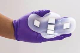
The MIT system starts from a probe roughly smaller than a deck of cards, built on a real-time 3D platform the group originally developed for breast-cancer detection. The clever part is the array geometry. Rather than packing a full grid of ultrasound elements, the team arranges the elements in the shape of an empty square, a hollow frame. That layout still captures enough spatial information to reconstruct a volume below the probe, but with far fewer elements than a conventional 3D array.
Fewer elements means less power and lower build cost, which is the practical lever that could move 3D ultrasound out of specialty departments. The probe moves its data off the device using a chirped data acquisition system, or cDAQ, a scheme that encodes the readout efficiently enough to stream in real time.
From there the pipeline takes a turn that will look familiar to anyone who has worked in game development or robotics simulation. The compressed voxel data streams into Unreal Engine, the same graphics engine behind a large share of modern games and an increasing amount of industrial digital-twin work. Unreal converts the voxel grid into a direct 3D rendering of the scanned structure, and the team reports no loss of information in that conversion. The user, wearing the headset, sees that rendering anchored over the physical object's actual position, an effect the researchers compare to X-ray vision. Tilt your head or step around the object and the view updates, so identifying an ambiguous structure becomes a matter of looking at it from another angle rather than re-running the scan in your imagination.
Using a general-purpose graphics engine as the rendering backend is a sensible engineering decision. Volumetric visualization, head-tracking, and spatial registration are problems that engines like Unreal already solve well, and leaning on that ecosystem keeps the medical team focused on the imaging physics rather than rebuilding a 3D renderer from scratch.
What the user study showed
The team tested AR-VIU with 18 participants split evenly between ultrasound experts, including sonographers and physicians, and people who had never touched an ultrasound. Each person ran identification tasks across four display conditions: standard 2D on a flat screen, 3D on a flat screen, AR in 2D, and the full AR-VIU 3D-in-AR setup.
The tasks were designed to mimic real clinical decisions. In one, participants identified an object such as a spring, a ball, or a screw embedded in gelatin inside an opaque container. In another, they used a pen to mark the location of a "tissue phantom," a gel engineered to mimic human tissue, standing in for the job of guiding a needle to the right spot during a biopsy.
AR-VIU improved identification and localization for everyone, but the effect on novices is the headline. With AR-VIU, first-time users performed nearly as well as experts. With traditional 2D imaging, the gap between the two groups was large, exactly what you would expect given the years of training the mental-reconstruction skill requires. "Overlaying images with the anatomy and providing 3D visual context makes ultrasound significantly easier for novices to understand," Viswanath says.
The subjective feedback split along predictable lines. Most novices preferred AR-VIU and said it made the tasks easier. "The 3D system imposes less brain drain, it's more intuitive, and it's easier to understand what is happening in the targeted region," Dagdeviren says. Many experts, on the other hand, preferred 2D, simply because it is what they trained on and trust. Those same experts still pointed to specific situations where they saw value in AR-VIU, such as guiding a biopsy needle or watching the heart wall move during echocardiography, cases where the 3D context adds something their trained intuition cannot easily supply.
Limitations and where it goes next
It is worth being precise about what this study does and does not establish. The objects were embedded in gelatin and tissue phantoms, not live patients, and the task set was identification and localization rather than diagnosis. Resolution is still a constraint the team is actively working on, and they plan additional testing to quantify the accuracy of the reconstruction against ground truth. Wearing a headset for an entire clinical shift raises ergonomic and workflow questions that a controlled study with short tasks does not answer.
The expert preference for 2D is also a real signal, not just habit. A trained sonographer has built a fast, reliable internal model, and a new visualization mode has to clear a high bar before it earns a place in an established workflow. The more compelling near-term case is the one the experts themselves named: specific high-stakes, spatially demanding procedures, plus training, where shortening the learning curve has obvious value.
That training angle connects to a broader pattern showing up across robotics and medical sensing. The expensive, scarce resource is often not the hardware but the human expertise needed to operate it. Systems that compress that expertise, whether through better visualization, learned assistance, or both, tend to expand where a tool can be used and who can use it. AR-VIU fits that pattern. By moving the 3D reconstruction out of the operator's head and into a rendering pipeline, it lowers the skill floor for a procedure that has historically demanded a great deal of it.
The work was funded by the MIT Media Lab Consortium, the National Science Foundation, an MIT HEALS graduate fellowship, and an MIT-Tata graduate fellowship. More on the group's broader program of wearable and conformable imaging devices is available through Dagdeviren's Conformable Decoders lab at the MIT Media Lab.
Comments
Please log in or register to join the discussion